


About Us
Executive Editor:Publishing house "Academy of Natural History"
Editorial Board:
Asgarov S. (Azerbaijan), Alakbarov M. (Azerbaijan), Aliev Z. (Azerbaijan), Babayev N. (Uzbekistan), Chiladze G. (Georgia), Datskovsky I. (Israel), Garbuz I. (Moldova), Gleizer S. (Germany), Ershina A. (Kazakhstan), Kobzev D. (Switzerland), Kohl O. (Germany), Ktshanyan M. (Armenia), Lande D. (Ukraine), Ledvanov M. (Russia), Makats V. (Ukraine), Miletic L. (Serbia), Moskovkin V. (Ukraine), Murzagaliyeva A. (Kazakhstan), Novikov A. (Ukraine), Rahimov R. (Uzbekistan), Romanchuk A. (Ukraine), Shamshiev B. (Kyrgyzstan), Usheva M. (Bulgaria), Vasileva M. (Bulgar).


Medical sciences
 PDF
PDFSummary: The application of new medical visual technologies, including ultrasound that occupies a leading position in the diagnosis of pneumonia and its complications in children is one of the promising trends in pediatrics. There are still inconsistencies in the application of ultrasound diagnostics in the diagnosis of chest disease, including pneumonia. But, nowadays it is century of the non-ionizing, fast, available diagnostic methods, which is sonography. This is especially actual in pediatrics.
Keywords: sonography, pneumonia, lungs, inflammation, pediatrics.
Relevance. Pneumonia diagnostics in children is one of the most actual problems in pediatrics. Currently, pneumonia holds the leading place in mortality causes structure after cardiovascular pathology, cancer diseases, cerebrovascular pathology and chronic obstructive pulmonary disease. [3].
Despite the variety of methods of diagnosis and treatment, mortality from pneumonia remains high. According to WHO, pneumonia kills approximately 1,1 million children each year - more than AIDS, malaria and measles together [8].
According to UNICEF, the mortality from pneumonia among children under the age of 5 years is 20% in developing countries and in the world 19%. In South Asia and sub-Saharan Africa is 21%, the Middle East and North Africa 15%, in the Pacific and East Asia, 15% in Latin America and of the Caribbean islands 14% in Central and Eastern Europe as well as in CIS countries 13% [4].
In Uzbekistan, the incidence of pneumonia among children continues to be high and does not tend to decrease. In 2012, the figures of respiratory diseases were - 17105.94, and among children -48,156.46 cases per 100 thousand of population; In 2013, these figures have increased considerably, 17863.57 and 50127.98 respectively [7]. If in this case we consider that the level of hospitalization with pneumonia complicated forms to remain high both in Uzbekistan and in other countries, imperfection of timely diagnosis and prevention of this disease becomes quite obvious. The researchers believe that the main efforts to reduce mortality from pneumonia should be focused on the development of algorithms for early diagnosis in order to select a rational therapy.
The application of new medical visual technologies, including sonography that occupies a leading position in the diagnosis of pneumonia and its complications in children is one of the promising trends in pediatrics [1, 2, 9].
Nowadays sonography is the most used method in the “medicine world”. Because it is very fast, available, nonionizing and mobile diagnostic method.
Materials and methods. This paper is based on the results of a complex standard examination of 80 children at the age of 1 - 18 years (50 children with pneumonia and 30 healthy children of the control group). Integrated clinical-laboratory and instrumental examination included a detailed medical history, physical examination blood tests, a comprehensive ultrasound scan of lungs with Doppler, X-ray examinations in clinic of Tashkent Pediatric Medical Institute.
The aim of the study is to optimize the diagnosis of pneumonia and its complications in children by the use of ultrasound scanning in system of complex examination of patients.
Results and discussions
50 children had various of clinical forms of acute pneumonia and pulmonary complications, and for 30 children pathology of the lung have not been identified (control group). There were 28 boys, and 22 girls. The distribution of examined children by age, in accordance with the classification of A.A Baranova (2007).
30 healthy children were studied especially in relation to ultrasound anatomy of the chest wall, pleura, lungs, mediastinum and diaphragm.
The distribution of examined children relative to clinical pneumonia in accordance with the classification of the clinical forms of bronchopulmonary diseases in children adopted in Moscow symposium on improving the classification of non-specific lung diseases in children (1995) is shown in Fig. 1.
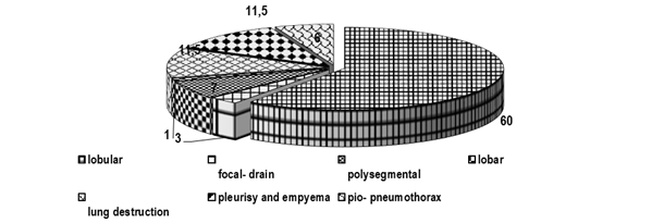
Fig. 1. Clinical forms of pneumonia and pulmonary complications in children in percentage (n = 50).
44 (88%) of children enrolled in the clinic with different clinical forms of uncomplicated pneumonia, and 6 (12%) patients had the disease associated with pulmonary complications.
44 (71.3%) of 50 patients with various forms of pneumonia had uncomplicated pneumonia.
In the vast majority of cases lung lesion was bilateral - 26 (59%) patients. 15 (34%) of children had right-sided pneumonia and 3 (6.8%) - left-sided pneumonia. Plan radiography of chest determined darkening lung fields for 50 patients, the size of the fields depended on the prevalence of lesions. In order to determine its nature ultrasound of the chest was performed to patients.
Aerial lung is impassable for the ultrasound waves, and airless lung sections are clearly visualized with echography. Alveolar gas fully reflects ultrasound, and echographic visualization of unaltered pulmonary parenchyma is impossible. However, in case of inflammation, alveoli are filled with exudate, there occurs edema of interstitial tissue, there is a strengthening of blood filling. Due to this, the lung tissue becomes airless and accessible to ultrasound.
Clinical echografically in 30 (81.8%) were diagnosed with focal, in 2 (3%) focal-drain, in 6 (11.5%) polysegmental, and 4 (7%) equity forms of pneumonia.
In case of echographic study, 50 patients had inflammatory infiltrate lung parenchymal visualized as a hypoechoic area with clear smooth outer contours. Pulmonary contours were indistinct due to the bordering air infiltrate the pulmonary parenchyma. The form of pneumonic focus was different. In the case of focal pneumonia, it was rounded - 4 cases, but more often irregular in shape - 33 patients, airless pockets of low echogenicity were noticed with the focal-discharge forms, which merged with each other (2 patients (4%)), in case of polysegmental pneumonia there was a pyramidal shape with base converted to the pleura (2 patients (4%)), in the case of lobar pneumonia, it repeated the form of a share (3 patients (6%)).
2 (4%) of patients with lung infiltrate had fluid accumulation on the affected side, which was detected by ultrasound in the pleural cavity in a small amount: while there were separation pleural sheets less than 15 mm.
In 35 (70%) cases, airless pockets of round and irregular shape were echografically visualized, they had low echogenicity with echogenic broken, radially reaching the lung central departments with linear structures with reverberations and echo shadow behind them (air-filled bronchi) and with hyperechoic ramifications of the bronchi in the form of "sprigs".
19 (38%) of patients with polysegmental pneumonia, under dynamic sonographic control in pulmonary infiltrates within one or more of the shares, had several anehogennoe small inclusions, the size of which does not exceed 2-4 mm. (Forming pockets of destruction of lung tissue). This echographic picture correlated with the negative dynamics of the clinical process. Subsequent ultrasound monitoring data foci gradually decreased in size and number, and then disappeared.
All 46 (92%) of the children, on the background of ongoing complex intensive therapy, exercised regular dynamic echographic control of the state and pneumonic hearth of the pleural cavity.
38 (76.0%) of 50 children, with dynamic ultrasound echographic recovery, observed following criteria: pulmonary infiltrates gradually decreased in size, its contours become indistinct, blurred, the number of displayable bronchial tubes filled with air, - i.e. pneumatization lung tissue restored. Pneumatization occurred in the direction from the root to the lung periphery. The amount of fluid in the pleural cavity on the side of the lesion gradually decreased in size, and in the case of recovery effusion was not visualized.
The diagnostic accuracy of sonography in the diagnosis of uncomplicated pneumonia, according to our data was 96.7 + 1.3%, while in case of plan radiography - 80.2 ± 3.0%.
Studies have shown that echography in the diagnosis of inflammatory infiltrates with uncomplicated pneumonia in children is superior to plain radiography informativeness. Echography made it possible to determine the nature of the blackout during imaging and detection of inflammatory infiltrate - to conduct monitoring during the treatment, without the use of repeated X-ray examinations, which is relevant in pediatric radiology.
2. Fazylov A.A., Vasiliev A., Olhova E.B., Yusupalieva G.A., Ultrasound diagnosis in pediatric practice. – Tashkent, 2014.
3. Pneumonia / Newsletter -Geneva: World Health Organization, 2014. –Vol.331. - 3.
4. Pneumonia - The forgotten killer of children. World Health Organization / UNICEF, 2006: 44p.
5. Radiation diagnostics in pediatrics. -Moscow, 2009.
6. Safonov D.V., Shakhov B.E., Ultrasound diagnosis of inflammatory lung diseases. – Moscow, 2011.
7. Statistical materials on the activities of health care institutions of the Republic of Uzbekistan, 2013.
8. World Health statistics 2014. Geneva: World Health Organization, 2014. -Vol. 45: 113.
9. Yusupalieva G.A., Ultrasound diagnosis of pneumonia and its complications in children.- Tashkent, 2009.
Yusupalieva G., Vaxidova N., Abzalov A. INFORMATIVE VALUE OF SONOGRAPHY IN CHILDREN WITH PNEUMONIA. International Journal Of Applied And Fundamental Research. – 2016. – № 6 –
URL: www.science-sd.com/468-25101 (25.07.2026).