


About Us
Executive Editor:Publishing house "Academy of Natural History"
Editorial Board:
Asgarov S. (Azerbaijan), Alakbarov M. (Azerbaijan), Aliev Z. (Azerbaijan), Babayev N. (Uzbekistan), Chiladze G. (Georgia), Datskovsky I. (Israel), Garbuz I. (Moldova), Gleizer S. (Germany), Ershina A. (Kazakhstan), Kobzev D. (Switzerland), Kohl O. (Germany), Ktshanyan M. (Armenia), Lande D. (Ukraine), Ledvanov M. (Russia), Makats V. (Ukraine), Miletic L. (Serbia), Moskovkin V. (Ukraine), Murzagaliyeva A. (Kazakhstan), Novikov A. (Ukraine), Rahimov R. (Uzbekistan), Romanchuk A. (Ukraine), Shamshiev B. (Kyrgyzstan), Usheva M. (Bulgaria), Vasileva M. (Bulgar).


 PDF
PDFUp-to-day the role of neutrophils in mechanisms of development of kidney pathology is discussed [1, 2, 3]. In this aspect the great interest represents the research of metabolic status of neutrophils in the blood of patients with chronic diseases of kidneys (CDK). Teziography is one of the methods, which can help getting the integrated characteristic of metabolic processes.
The purpose of the real research was studying of morphotypes of leucocyte´s lysates received from blood of patients with chronic diseases of kidneys.
PATIENTS AND METHODS: 43 patients (29 females and 14 males, age 17-52 years) were examined; they divided in to three groups. In first group were patients with tubulopathy (TP) like chronic pyelonephritis in remission (without urinal infection). At patients of this group at ultrasonic investigation were observed the deformation of pyelocaliceal system of kidneys. Clinically the pyelonephritis proceeded with a small amount of signs. The patients with interstitial kidney pathology were included in second group. In the anamnesis at these patients was long use of medicines or toxicants. In urine tests on concentration was marked the decrease of relative density. Third group included the patients with glomerulopathy. Presence of glomerulonephritis is verified by characteristic urinary sediment and pathomorphologic picture at the nephrobiopsy.
Leucocytes were excreted from peripheral blood, using a method, offered by M.Z. Fedorova and N.V. Levin [4]. Leucocytes were destroyed by triple freezing and thawing. Volume of lysate (100 mkl) was put on the pure and fat-free slide plate. After drying the received facias were scanned and analyzed.
RESULTS AND DISCUSSION:
The typical theziogram of leucocyte´s lysates of healthy persons is represented on fig. 1.
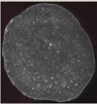
Figure 1. Theziogram of leucocyte´s lysates of healthy person
Crosswise pseudo-crystalline structures of medium dimensions were observed in the central zone of leukocyte facias of healthy donors. Intermediate zone has been presented by long crystals located radially, crosswise crystals and small crystals with abnormal form. In fringe zone there were single long crystals.
The typical theziogram of leucocyte´s lysates of the patient with intersticial nephritis (IN) is represented on fig. 2.
The theziograms of leucocyte´s lysates of patients with IN were characterized by presence of long crystals located in intermediate zone of facia with direction of crystal growth from centre to periphery. In direction to fringe zone the crystals were branched, forming the crystals of 2-nd and 3-rd orders. In the central zone of facia was observed a small region with pseudo-crystals located in type of scutal formations and generated in type of cruciate structures.
The typical theziogram of leucocyte´s lysates of patient with a chronic pyelonephritis (CPN) is represented on fig. 3.
The theziograms of leucocyte´s lysates of patients with CPN were characterized by presence of large scutal structures, formed by crystals, growing crosswisely. These structures were present in the most part of the facias, where was free only fringe zone. Fringe zone was characterized by presence of less amount of chaotically located small crystals.
The typical theziogram of leucocyte´s lysates of patient with glomerulonephritis (GN) is represented on fig. 4.
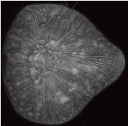
Figure 2. Theziogram of leucocyte´s lysates of the patient with IN
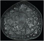
Figure 3. Theziogram of leucocyte´s lysates of patient with CPN.
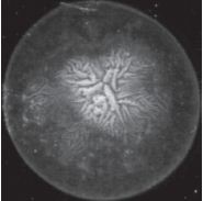
Figure 4. Theziogram of leucocyte´s lysates of patient with GN
The theziograms of leucocyte´s lysates of patients with GN were characterized by presence of small dendrite crystalline structures located in the central zone of facia. The intermediate zone of facia was transparent, with rare small crystals. Crystalline growth in the fringe zone of facia was not observed.
According to our researches the structure-forming properties of neutrophils of patients with CDK are characterized by expressed metabolic infringements. The metabolic infringements directly depend on prevailing type of damage of kidney tissue. Neutrophil´s lysates of patients with glomerulonephritis has more elementary architectonic of theziographic structures then at patients with pyelonephritis the teziogramms of which are less organized. All three groups have general tendency in development of pathological process, what is characteristic for CDK, but each of them has peculiarities in of neutrophil´s metabolism. In our opinion, these peculiarities significantly can influence on behavior of leucocytes in the conditions of chronic inflammatory process, defining their contribution in progressing of irreversible changes in kidney tissue.
References
- Heinzelmann M., Mercer-Jones M.A., Passmore J.C. // American journal of kidney diseases. - 1999, vol. 34, n 2.- P. 384-399.
- Costa E., Rocha E. Rocha-Pereira P. et al. // Haematologica. - 2007 - 92 [suppl.2]: 290. Abstract 0776
- Jaber B. L., Perianayagam M. C, Balakrishnan V. S et al // Journal of Leukocyte Biology. - 2001.- 69.- P. 1006-1012.
- Fedorova M.Z., Levin V.N. // Clinical medicine. - 2000. - №8. - P. 35-38.
Muravleva L.Е., Molotov-Luchansky V.B., Kluyev D.A., Kussainova D.S., Tuleshova A.Zh., Abdrakhmanova Yu.E., Kolesnikova E.A. MORPHOTYPES OF NEUTROPHIL’S LYZATES OF BLOOD OF PATIENTS WITH KIDNEY DISEASES. International Journal Of Applied And Fundamental Research. – 2009. – № 2 –
URL: www.science-sd.com/385-23407 (30.10.2025).